Endoscopy is a non-invasive diagnostic and therapeutic procedure that has revolutionized the field of veterinary internal medicine. Through the use of cameras on the end of various length tube-like instruments (endoscopes), internal areas of the body can easily be examined without the need for surgery. There are different types of endoscopic procedures, each for a specific organ or organ system. In this blog, we discuss rhinoscopy which is endoscopy of the nasal cavity and nasopharynx.
What is Rhinoscopy?
Rhinoscopy is a specific type of endoscopy that, as for all forms of endoscopy, involves the use of an endoscope (in this case, rhinoscope), a thin light-emitting tube equipped with a miniature camera on the tip. The rhinoscope is gently inserted into the nasal passages, allowing visualization of the internal structures of the nose. The images are displayed on a high-definition monitor with the ability to view and record both pictures and video. This procedure can be performed using either a rigid or flexible rhinoscope, depending on what is being evaluated.
The rhinoscope is gently inserted into the nasal passages, allowing visualization of the internal structures of the nose.
Internal Structure of the Dog Nose
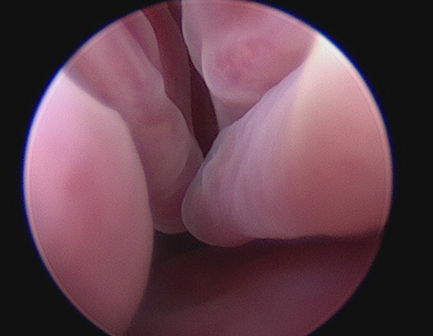
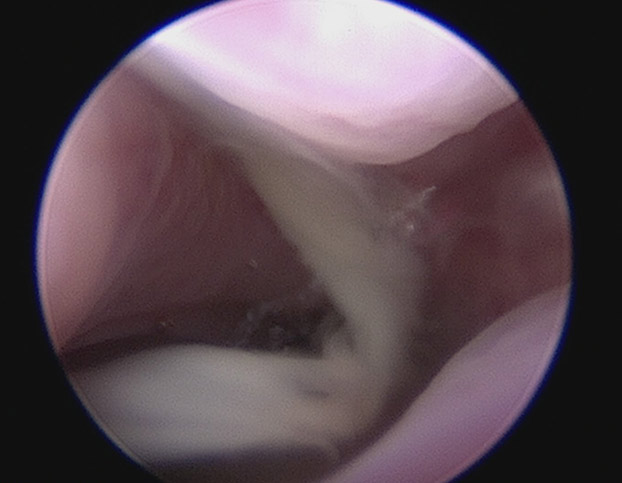
The inside of the dog nose is divided by the nasal septum into the left and right nasal cavities. Each nasal cavity is essentially filled with “turbinates” which are complex, complicated, branching pathways which significantly increase surface area allowing for temperature and humidity regulation of inspired air, added immunity against pathogens that may be inhaled and to provide the impressive smell capability of dogs. When a rhinoscopy is performed, the inside of the nasal cavities are not “open caverns”, but instead, a maze of “tunnels”, “shelves”, “outcroppings” and smaller passageways that inspired air travels around and through. This complex structure makes a thorough rhinoscopy where all areas have been viewed, a complicated procedure.
Rigid vs Flexible Rhinoscope
There are two different types of rhinoscopes used to perform a complete rhinoscopy procedure:
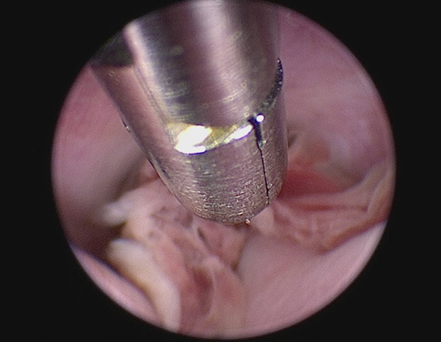
Rigid Rhinoscope: a short, slender, light-emitting medical-grade stainless steel “tube” with a camera embedded into the end. It is specifically designed to navigate the complex anatomy of the dog and cat nasal cavity.
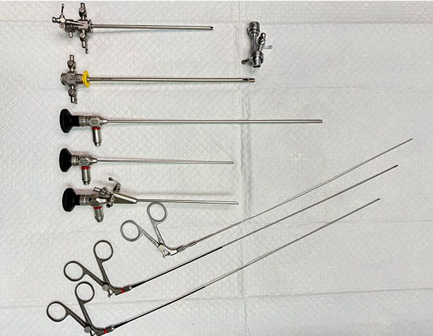

Flexible Rhinoscope: a longer tube-like light-emitting instrument equipped with a camera at its tip. Its flexibility allows the full visualization of the nasopharynx (space behind the nose, above the soft palate) by insertion in the mouth then flexing 180°. A flexible rhinoscope can be used to evaluate both the nose and throat at the same time, whereas a rigid rhinoscope can only be used to evaluate the nose.
Antegrade vs Retroflexed Rhinoscopy
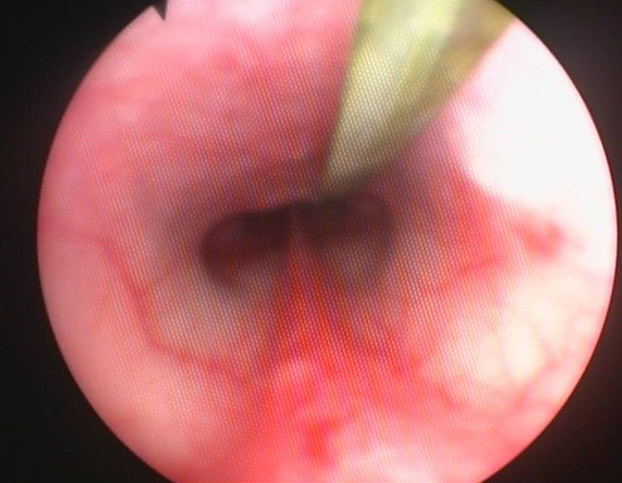
Antegrade Rhinoscopy: a rigid rhinoscope is inserted into each nostril and slowly and carefully maneuvered through and around the turbinates while advancing towards the back of the nasal cavity.
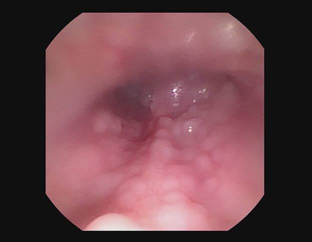
Retroflexed Rhinoscopy: a flexible rhinoscope is maneuvered into the nasopharynx (the area behind the nasal cavities and above the soft palate) via the mouth. The scope is then retroflexed 180° to examine the nasopharynx and the openings at the back of the nose. This method is particularly useful for examining the post-nasal structures and identifying retained foreign material and nasopharyngeal polyps.
The choice between rigid and flexible rhinoscopy, as well as antegrade and retroflexed rhinoscopy, depends on the specific requirements of the examination and what is being evaluated. We perform all of these for our patients undergoing rhinoscopy.
What Can and Cannot Be Seen?
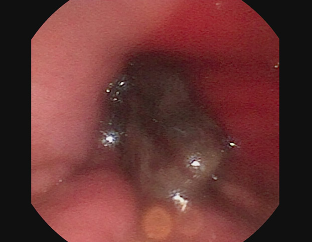
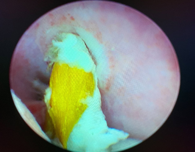
During a rhinoscopy, various structures within the nasal cavity can be seen, including the nasal septum, the turbinates, and the openings to the sinus passages. Abnormalities such as polyps, tumors, foreign material, fungal infection and areas of inflammation can be identified. However, rhinoscopy may not provide a full picture of the pet’s nasal health. For instance, it may not be able to detect conditions affecting the deeper structures of the nose or the sinuses. For these areas, advanced imaging such as CT scan may be required.
Reasons to Perform a Rhinoscopy
Rhinoscopy is typically indicated for pets exhibiting symptoms such as:
- Nasal Discharge
- Nosebleeds (Epistaxis)
- Facial or Nasal Deformity
- Nasal Obstruction
- Pawing at the Nose or Face
It can help diagnose a variety of conditions, including:
- Tumors (Cancer)
- Nasopharyngeal Polyps
- Fungal Infections
- Foreign Objects (and Removal)
- Nasopharyngeal Stenosis
- Idiopathic Lymphoplasmacytic Rhinitis
How Is a Rhinoscopy Performed?
The following outlines the steps in performing a complete rhinoscopy:
- General Anesthesia: Ensure the comfort and cooperation of the patient.
- Ventral Recumbency Position: Once anesthetized, your pet is positioned laying on their belly.
- Nasal Nerve Blocks: Reduce discomfort and the amount of general anesthesia needed, nerve blocks are performed to “numb” the nose.
- Buccal Mucosal Bleeding Time: Ensure your pet has normal ability to clot their blood.
- Retrograde Rhinoscopy: The nasopharynx is evaluated using a flexible rhinoscope. Any abnormal tissue is biopsied, and any foreign material is removed.
- Protect the Airway: The back of the oral cavity (pharynx) is tightly packed and occluded with gauze. This prevents aspiration of the saline (see next step) and nasal contents into the trachea and lungs.
- Antegrade Rhinoscopy: Using a rigid rhinoscope, both nasal passages are viewed via insertion into the nostrils. Saline solution is infused through the rhinoscope and into the nasal passages during this step to move any mucus or blood to provide a clear view.
- Nasal Flushing: The final step is flushing of the nasal passages with large amounts of saline solution. As the solution drains from the nose, it is strained through gauze to catch any abnormal tissue or undetected foreign material that may be flushed out.
- Anesthesia Recovery: The gauze in the pharynx is removed and the patient recovered from anesthesia.
What Pet Parents Need to Know Before the Procedure
Because your pet’s health is of the utmost importance, there are several important steps prior to performing a rhinoscopy:
- A minimum of a 12-hour fast — no solid food, water is always allowed and should not be withheld.
- A thorough physical examination is performed by a veterinarian to detect any new or unknown health issues that your pet may have.
- Recent pre-anesthetic blood tests are needed to ensure your pet is safe for anesthesia.
- If your pet has heart or lung concerns, pre-anesthesia EKG and thoracic X-rays may be important
What Pet Parents Need to Know After the Procedure
Rhinoscopy is an outpatient procedure, and your pet will be able to go home 1-2 hours after completion of the procedure. Your pet may be groggy or sleepy for several hours after returning home. It is not uncommon to see an intermittent (especially brought on by sneezing or exercise) slightly bloody nasal discharge for the first 12 hours after going home — this is normal and poses no concerns; restricting vigorous exercise during these first 12 hours may reduce this. Your pet may eat a small meal when they return home. By the following day, there should be no lingering effects of the procedure.
Your pet may be groggy or sleepy for several hours after returning home.
Rhinoscopy vs Surgery
Rhinoscopy offers several advantages over traditional surgical methods. It is a minimally invasive procedure that allows for a detailed examination of the nasal passages without the need for a surgical incision. This can result in less discomfort, a quicker recovery time and no facial scars or disfigurement. However, rhinoscopy does have the limitations that large tumors or masses may not be able to be completely removed, but only biopsied (sampled).
Your Pet’s Safety and Your Confidence is Our Priority
In conclusion, rhinoscopy is a valuable tool in veterinary medicine, providing a minimally invasive method for diagnosing and treating a variety of nasal conditions. However, like any medical procedure, it has its limitations and risks. We understand that you worry about your furry family member, and we will spend as much time discussing all aspects of the procedure with you prior to the procedure to ensure that you are comfortable and confident. Please contact us if you have questions about rhinoscopy or wonder if your pet would benefit from this procedure.
Author:
James Woods DVM, MS, DACVIM (SAIM)
Ph: (912) 721-6410
Contact Us