Endoscopy is a non-invasive testing and treatment procedure that uses various length light-emitting tube-like instruments (endoscopes) to evaluate internal areas of the body, all without the need for surgery. Endoscopes have a miniature camera embedded into the tip which allows a non-invasive view deep inside the body. There are different types of endoscopic procedures, each for a specific organ or body part. In this blog, we discuss upper gastrointestinal endoscopy which is endoscopy of the upper portion of the digestive tract.
What is Upper GI Endoscopy?
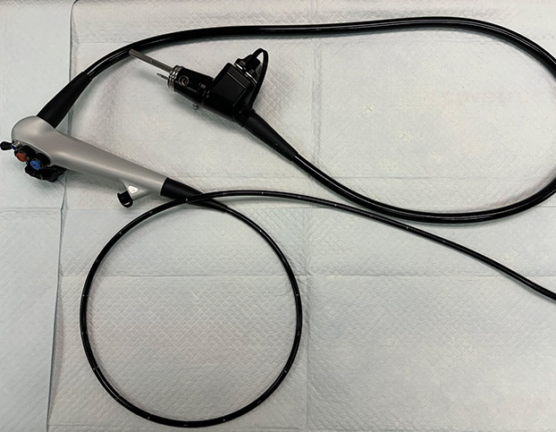
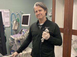
Upper GI Endoscopy (esophagogastroduodenoscopy) is a specific type of endoscopy that evaluates the first part of the digestive tract including the esophagus, stomach and duodenum (beginning of the small intestine). The endoscope (gastroscope) is 120 cm in length, and flexible to maneuver around corners. The internal details of the esophagus, stomach and duodenum are displayed in real time on a high-definition monitor, and both still photos and videos can be recorded.
The endoscope (gastroscope) is 120 cm in length, and flexible to maneuver around corners.
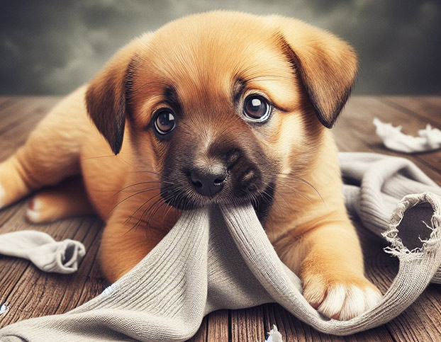
Why Your Pet Might Need an Upper GI Endoscopy
- Evaluate unexplained symptoms, such as:
- Drooling
- Not eating
- Painful swallowing
- Inability to swallow
- Frequent vomiting or regurgitating
- Weight loss
- Diarrhea or black stools
- Diagnose a variety of diseases, such as:
- Polyps, tumors and cancers of the esophagus, stomach and duodenum
- Ulcers of the esophagus, stomach and duodenum
- Esophageal strictures
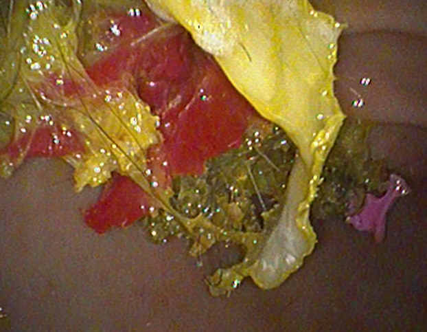
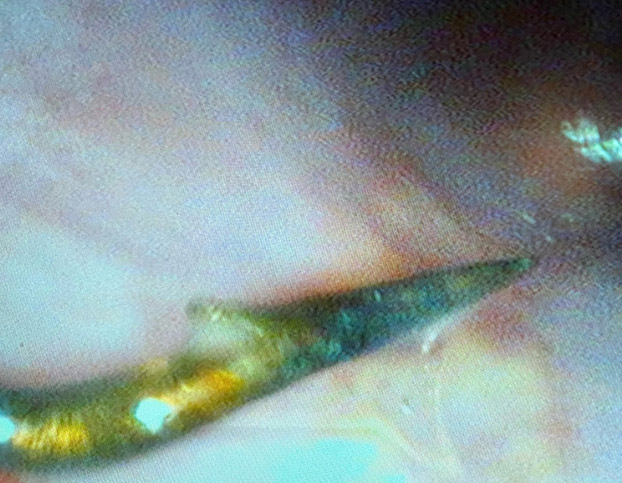
- Retained foreign material or lodged non-digestible food items
- Inflammatory bowel disease
- Hiatal hernia
3.Treat certain conditions, such as:
- Balloon dilation of esophageal strictures
- Removal of swallowed items that are lodged in the upper digestive tract
- Assist in the placement of feeding tubes, such as esophageal feeding tube and gastrostomy tube.
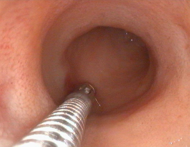
4. During an upper GI endoscopy, small samples (biopsies) of any tissue that appears abnormal (swollen, inflamed, cancer) can be taken directly through the endoscope without surgery. Any samples obtained during the procedure are sent to a veterinary pathology laboratory for analysis (histopathology).
What Pet Parents Need to Know Before the Procedure
Because your pet’s health is of the utmost importance, there are several important steps prior to performing an upper GI endoscopy:
- A minimum of a 12-hour fast — no solid food, water is always allowed and should not be withheld.
- A thorough physical examination is performed by a veterinarian to detect any new or unknown health issues that your pet may have.
- Recent pre-anesthetic blood tests are needed to ensure your pet is safe for anesthesia.
- If your pet has heart or respiratory concerns, pre-anesthesia EKG and thoracic X-rays may be important
How is an Upper GI Endoscopy performed?
The following outlines the steps in performing a complete upper GI endoscopy:
- General Anesthesia: Ensure comfort and cooperation. Your pet is continuously monitored while under anesthesia by a veterinarian and a registered veterinary technician (RVT). Oxygen levels, breathing, heart rate, EKG, blood pressure and temperature are continuously monitored. Intravenous (IV) fluids are given throughout the procedure to support blood pressure and the kidneys.
- Left Lateral Recumbency Position: Once anesthetized, your pet is positioned laying on their left side.
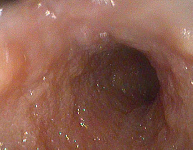
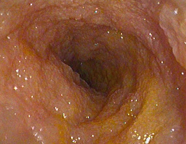
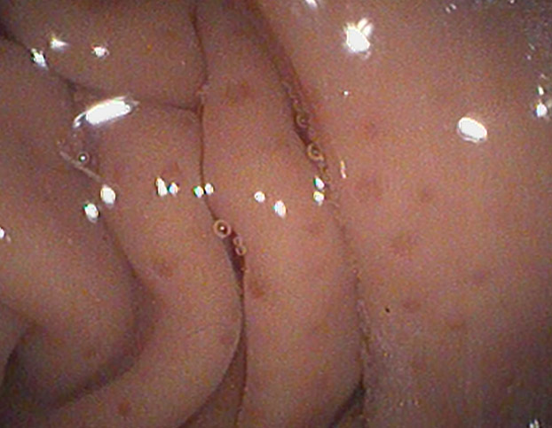
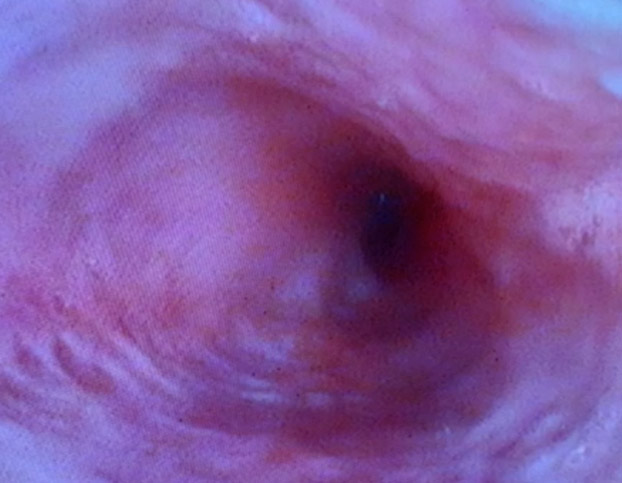
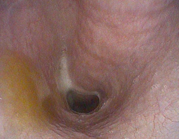
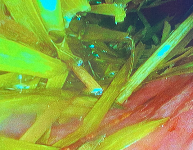
- Esophagus Evaluation: The endoscope is carefully inserted into the mouth and gently directed into the esophagus. The esophagus is evaluated for ulcers, gastric flux (GERD), narrowing (strictures). Any retained swallowed foreign material is removed and esophageal strictures can be dilated and “stretched” open.
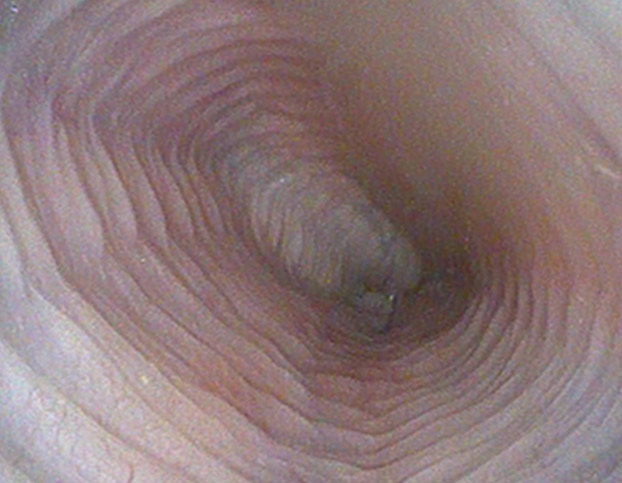
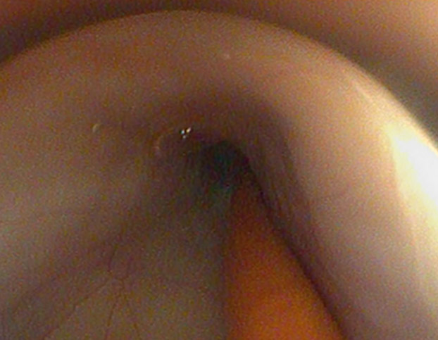
- Stomach Evaluation: The endoscope is slowly advanced through the lower esophageal sphincter (valve between esophagus and stomach) into the stomach. The stomach is insufflated with air through the endoscope so that the endoscope can be safely advanced, and to allow the entire stomach to be seen. Any foreign material is removed and abnormal tissue biopsied.
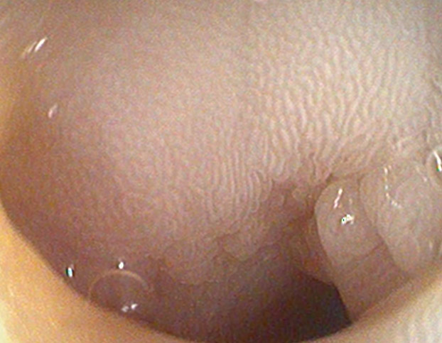
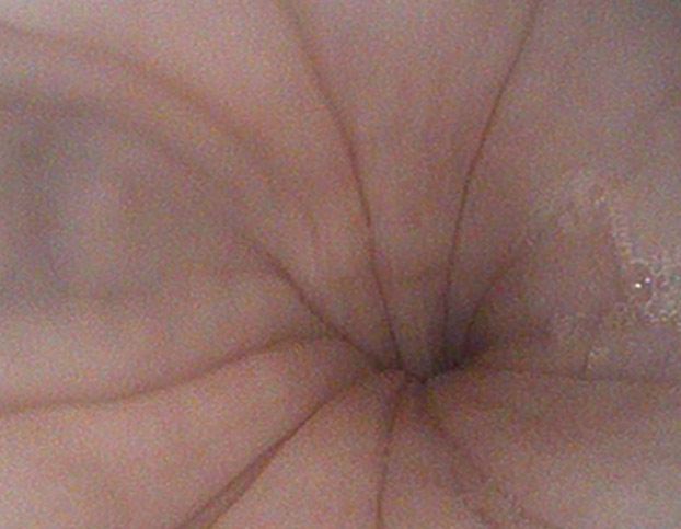
- Duodenum Evaluation: The endoscope is gently advanced through the pylorus (valve between the stomach and duodenum) into the first part of the small intestine. The small intestine is insufflated with air through the endoscope to improve visualization and facilitate its safe maneuvering. The color and texture of the lining of the duodenum is visualized and any abnormal areas biopsied.
- Recovery from Anesthesia: Any insufflated air remaining in the duodenum and stomach is suctioned prior to removing the endoscope and your pet being awakened from anesthesia.
What Pet Parents Need to Know After the Procedure
- Upper GI endoscopy is an outpatient procedure and your pet will go home the same day.
- The procedure is not painful, and pain relievers are generally not needed.
- Your pet may be groggy or tired for 6-8 hours — this is normal. Your pet should stay in the house and be observed during this time; they may need help on stairs and getting in/out of the car.
- Your pet can eat a small meal after arriving home.
- Strenuous exercise should be avoided for the first 12 hours after the procedure. It is important to allow any remaining insufflated air to be naturally expelled to avoid stomach bloat.
- Your pet may have a very small amount of dark (black) bowel movement after the procedure. This is normal and is caused by minor bleeding at the time any biopsies were taken.
- The following day, normal activity and lifestyle may resume.
- Results of any biopsies or other samples can take 5-7 days and we will call you to discuss.
- If you have any concerns or questions after returning home with your pet, please contact us for advice.
Upper GI Endoscopy vs. Surgery
Endoscopy offers many benefits over surgery, including:
- Non-invasive — no incisions, cutting, scarring
- Short recovery time
- Minimal discomfort
- Outpatient procedure, no lengthy or overnight hospital stays
Endoscopy is our preferred method over surgery for evaluating:
- Visual inspection of the esophagus, stomach and first part of the small intestine
- Obtaining diagnostic samples (biopsies) of the upper GI tract
- Eliminating foreign material ingestion by endoscopic retrieval
- Correction of esophageal strictures with balloon dilation
- Insertion and visual inspection of a variety of feeding tubes to assist patients who cannot eat on their own
Your Pet’s Safety and Your Confidence is Our Priority
In conclusion, upper GI endoscopy is a valuable tool in veterinary medicine, providing a minimally invasive method for diagnosing and treating a variety of upper digestive conditions. However, like any medical procedure, it has its limitations and risks. We understand that you worry about your furry family member, and we will spend as much time discussing all aspects of the procedure with you prior to the procedure to ensure that you are comfortable and confident. Please contact us if you have questions about upper GI endoscopy, or wonder if your pet would benefit from this procedure.
Author:
James Woods DVM, MS, DACVIM (SAIM)
Ph: (912) 721-6410
Contact Us