If you’ve been told that your dog or cat needs an ultrasound, you probably have questions. How does it work? What does it show? Why does my pet need an ultrasound when they already had X-rays? As the most experienced veterinary internal medicine specialty practice along the Georgia coast, you’ve come to the right place to find the answers to all your ultrasound questions. We’ve performed over 15,000 ultrasound procedures, and there is little we haven’t seen.
How does ultrasound work?
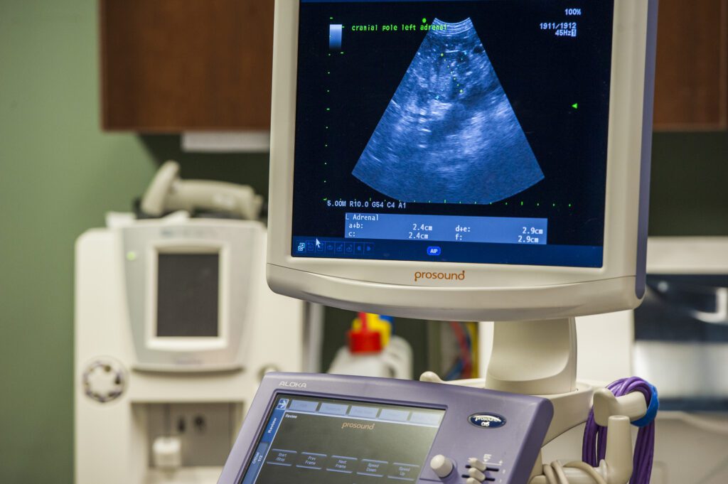
Using a probe (aka “transducer”) that is gently pressed against the skin, high-frequency sound waves are beamed into the body, and those sound waves rebound or bounce off body parts and return to the transducer. Using calculations including the speed of sound and the time that the sound waves take to return to the transducer, a computer is able to produce images of what the sound waves interacted with. The sound waves do not cause pain or discomfort, and there are no lasting effects.
What is Doppler ultrasound?
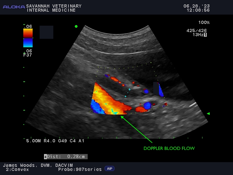
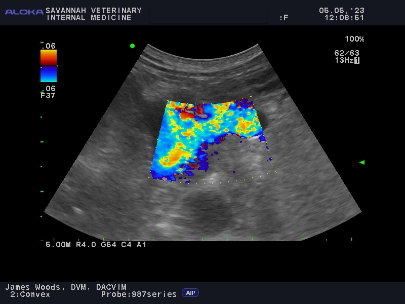
Doppler ultrasound, also using high-frequency sound waves, measures the flow (velocity, direction) of blood through blood vessels (veins, arteries, capillaries) and the heart. It is used to determine if blood is flowing normally and whether there are any blood clots or other blockages. Doppler ultrasound is very useful in almost all patients that we see for an ultrasound.
Do pets cooperate for their ultrasound? Do pets need anesthesia to have an ultrasound performed?
Most pets are very cooperative and sedation is rarely needed. Our veterinary technicians will gently hold your pet while they lie on their back in a foam, padded V-shaped trough. Since the transducer needs to make contact with the skin, fur overlying the area to be viewed will be shaved and a water-based gel applied to the skin. Our doctors then perform the ultrasound procedure — our technicians do not perform these procedures, these are performed directly by the specialist. Depending on the complexity of your pet’s health issues and the number of areas that need to be scanned, the procedure can take up to 30 minutes in some patients. Ultrasound is performed in a darkened room and many patients fall asleep while it is being performed!
My pet isn’t pregnant, why do they need an ultrasound?
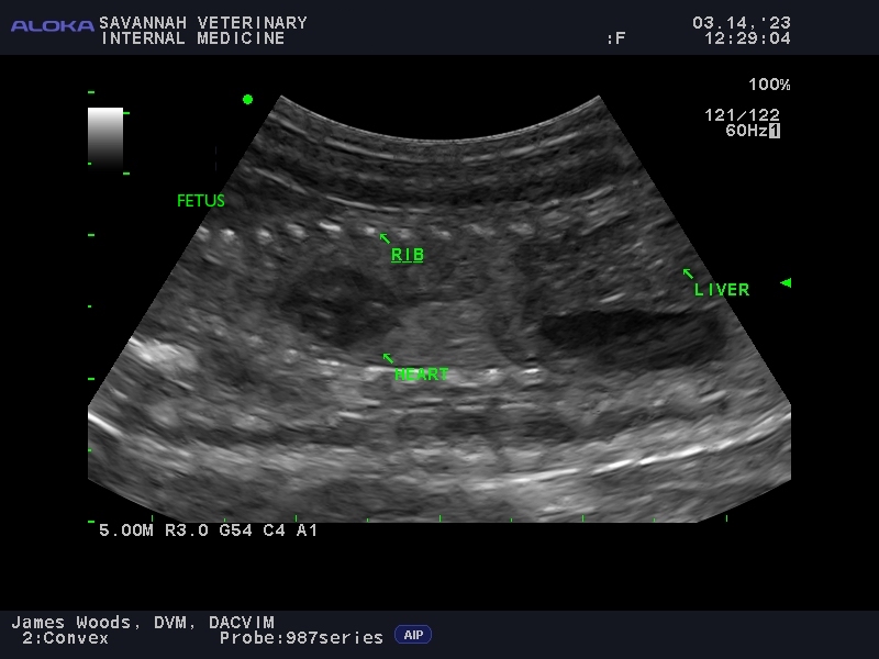
Many people’s familiarity with the word “ultrasound” or “sonogram” starts and ends with their experiences with pregnancy. Although it is useful for viewing fetuses (puppies and kittens included!), its usefulness extends way beyond that assisting in diagnosing and managing diseases. Diseases that can be “seen” with ultrasound range from simple, to complex, to life-threatening. However, rest assured that just because it has been recommended that your pet have an ultrasound, this does not mean anything serious will be found.
My pet had X-rays, why did they need an ultrasound?
We often hear the incorrect assumption that an ultrasound is not needed because X-rays were already performed (or vice versa). Although both imaging tools — X-rays (aka radiographs) and ultrasound — “look inside the body” they do it in very different ways, and provide very different information. It’s like saying “I don’t need a car because I have a bicycle”. Both are able to get you from point A to point B, but they do it in very different ways and are used in much different situations.
X-rays produce a two-dimensional image highlighting the shape, number and density of organs, including bones; in fact, X-rays are exceptional at evaluating bones whereas ultrasound is poor at evaluating bones. Like X-rays, ultrasound also produces two-dimensional images and is also useful for evaluating the shape, number and density of organs, but it also provides information about subtle details of the inner architecture of organs, the presence of inflammation associated with organs, evaluating blood flow (through veins, arteries, the heart and other organs), locating small cancerous nodules and masses and for detecting small volumes of fluid surrounding organs. Both ultrasound and X-rays play important roles in the diagnosis and management of many internal medicine diseases and frequently these are used together to paint a clearer picture of your pet’s internal organs.
Exposure to X-rays can be harmful, will ultrasound harm my pet?
It is true that repeated exposure to X-rays can cause cancer. Ultrasound on the other hand is completely safe, no precautions are needed to be near it and the procedure will not hurt your pet or our staff assisting during the procedure.
Which common internal medicine diseases can ultrasound detect and diagnose?
Although there are hundreds and possibly even thousands of diseases and abnormalities ultrasound can detect, there are common ones that we see on a daily basis. Below we list a handful to show the usefulness of ultrasound in helping our patients.
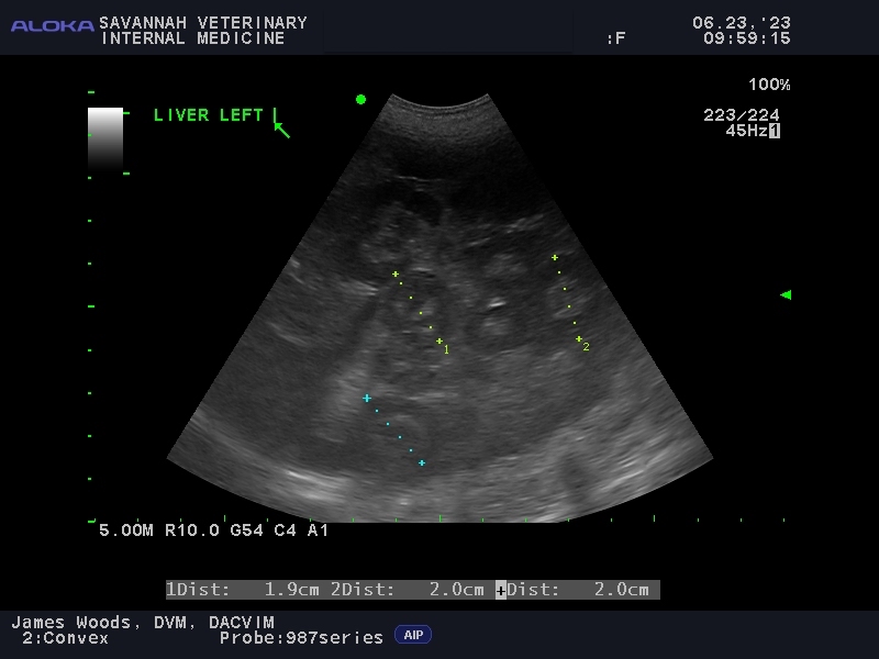
Liver
Many diseases and disorders of the liver are easily recognizable with ultrasound, including cancer, hormone-induced changes (diabetes mellitus, Cushing’s disease), congenital diseases (portosystemic shunt, microvascular dysplasia), and metabolic conditions (hepatocutaneous syndrome, hepatic lipidosis in cats).
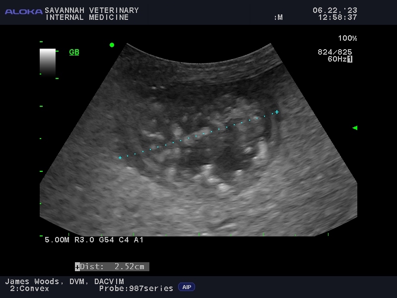
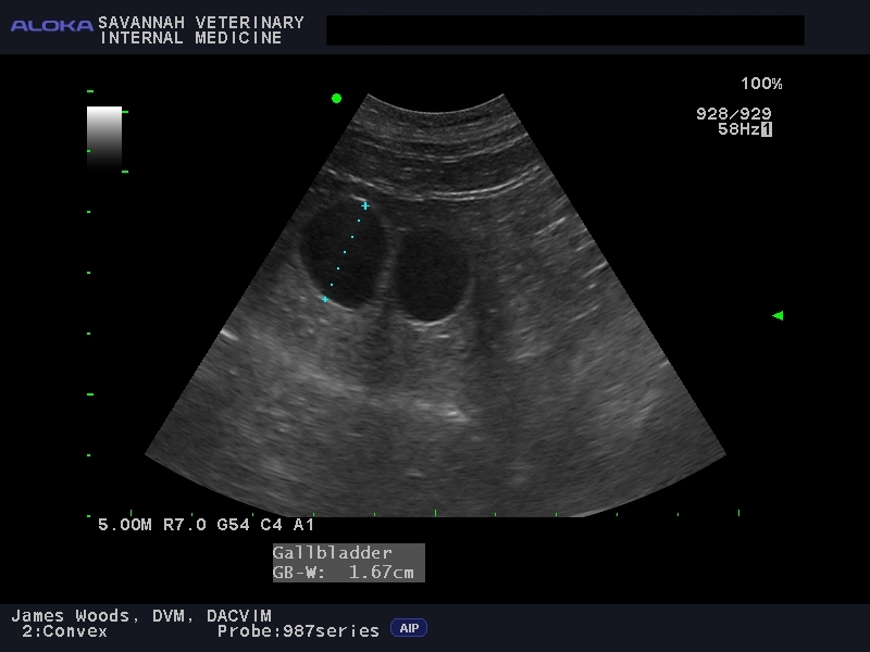
Gallbladder
Diseases and syndromes of the gallbladder and biliary ducts that are frequently detected with ultrasound include biliary mucocele, inflammation/infection (cholecystitis), stones (gallstones or stones in the bile ducts) and biliary tree rupture with bile leakage. Cats occasionally have an “extra” gallbladder (bilobed, duplex or complex) which does not cause illness or symptoms.
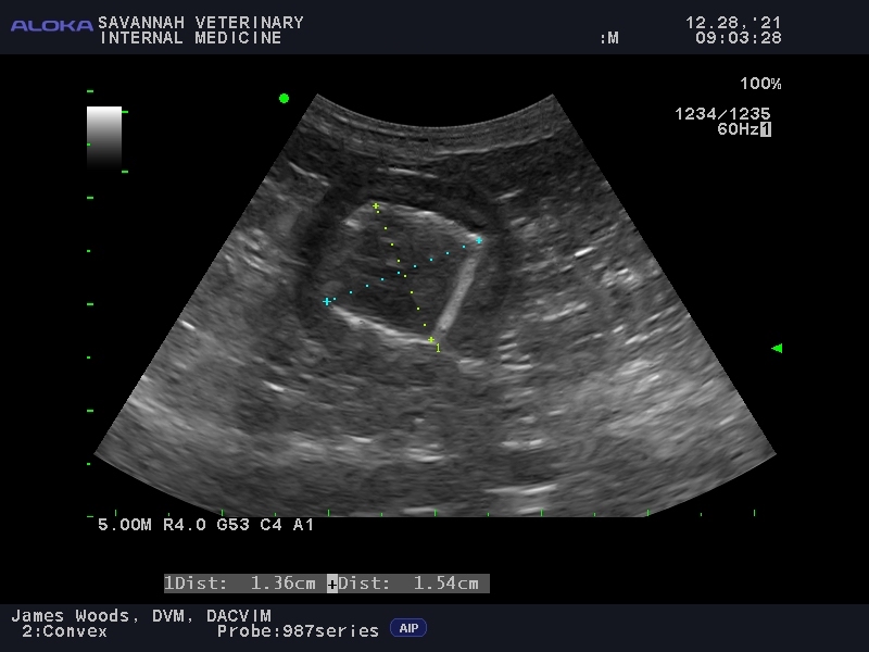
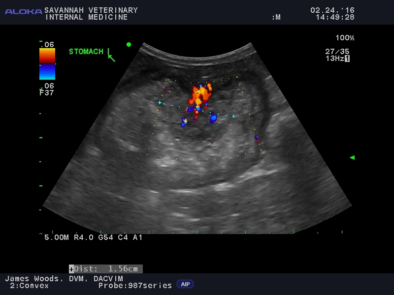
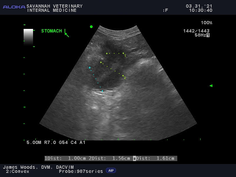
Stomach
A variety of stomach diseases and conditions are easily recognizable with ultrasound including stomach wall cancer, stomach wall infection and abscess, ulcers, retained foreign material and pyloric hypertrophy.
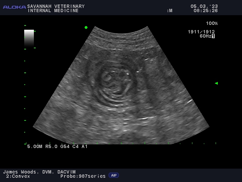
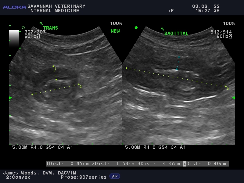
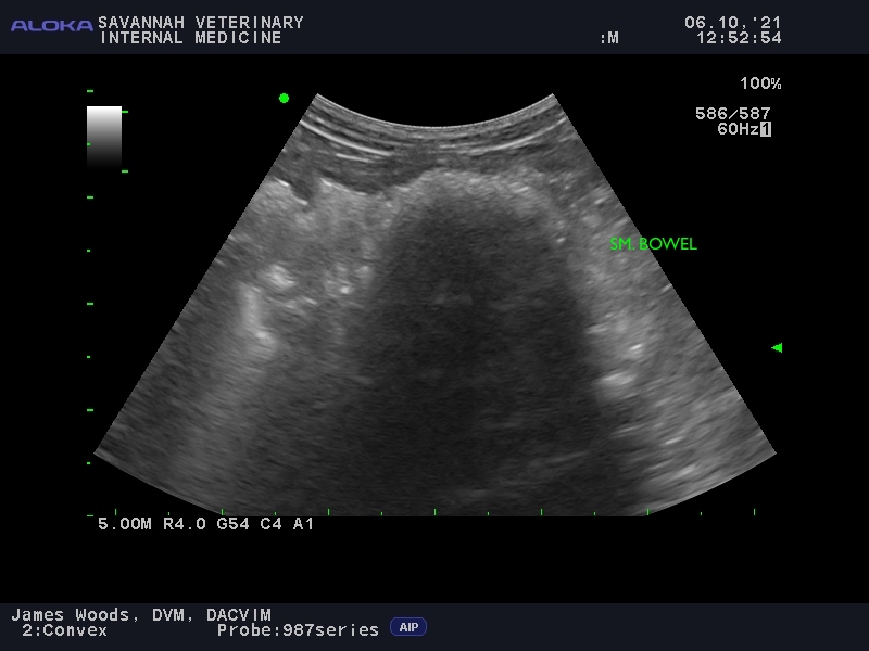
Intestinal tract
The intestinal tract includes the small intestines and the colon. Diseases affecting these organs that can be clearly visualized with ultrasound include inflammatory bowel disease, intestinal cancer, retained foreign material, intestinal obstruction from masses or foreign objects that have been swallowed and intussusception (telescoping of the intestines into an adjacent segment resulting in an obstruction).
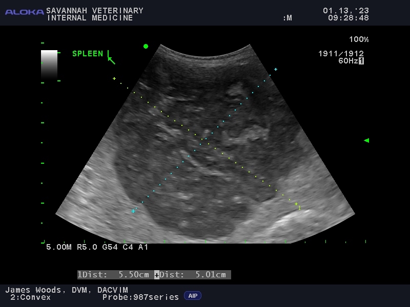
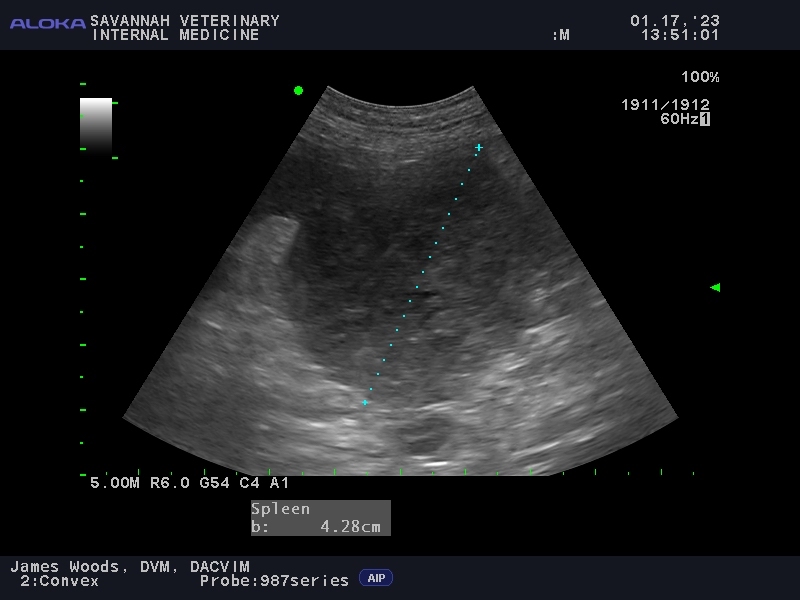
Spleen
The spleen is an immune organ important for preventing infections and cancers. Splenic diseases easily seen with ultrasound (and frequently invisible to X-rays) include cancerous or benign (hyperplastic, myelolipoma) masses and scarring.

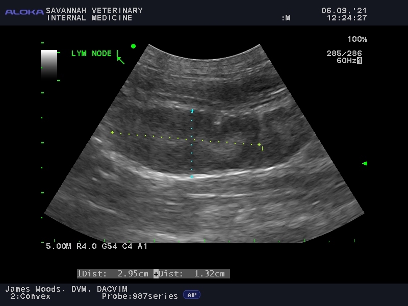
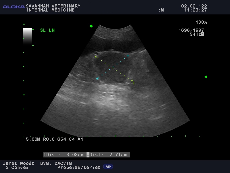
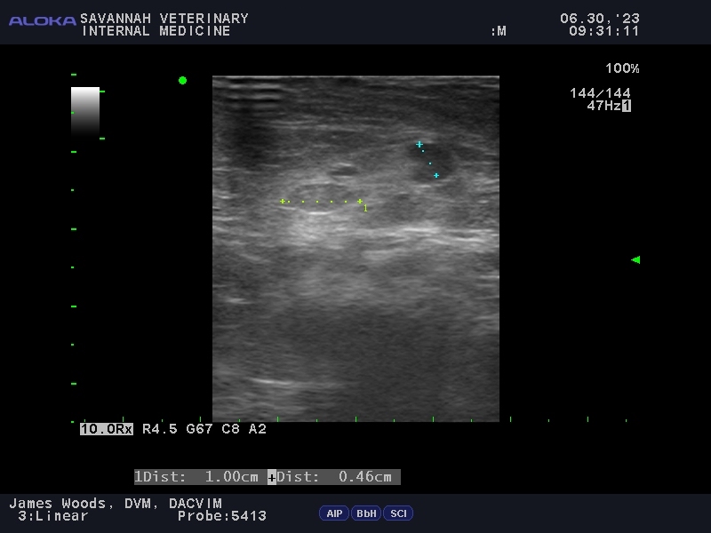
Lymph nodes
There are hundreds of lymph nodes throughout your dog and cat’s body, many of which are contained within the chest and abdominal cavities. Lymph nodes are part of the important lymphatic system, which is responsible for immune surveillance and protection from infection and cancer. Unlike X-rays, ultrasound excels at detecting even the smallest changes to lymph nodes.
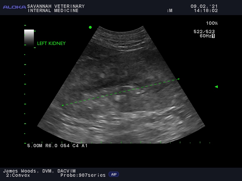
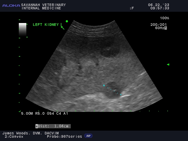
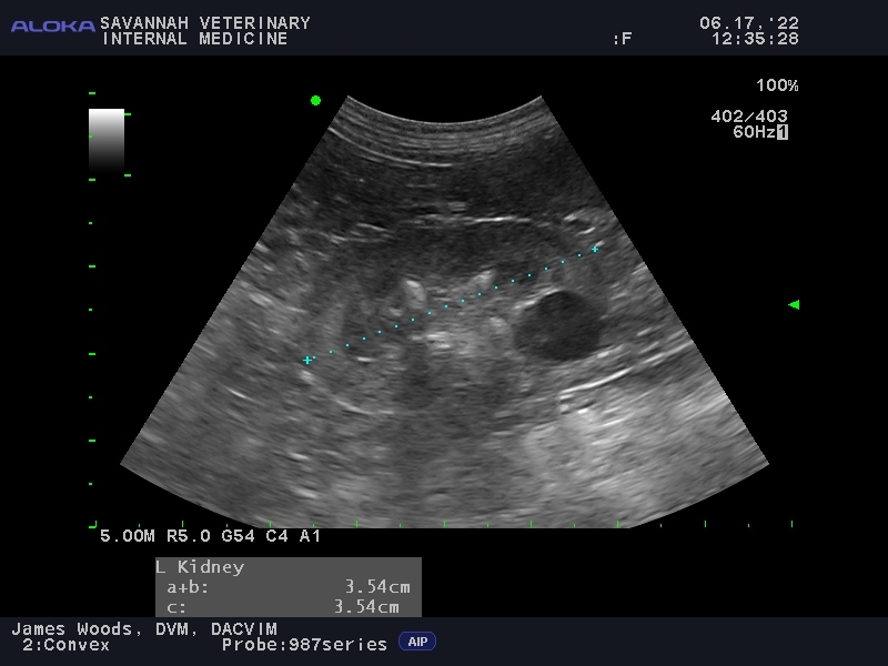
Kidneys
Although X-rays can determine the presence, size and shape of kidneys, ultrasound is able to provide additional information regarding the internal architecture of the kidneys, and whether chronic kidney disease (CKD), congenital kidney disease (renal dysplasia) or kidney cancer is present. Using Doppler ultrasound, the blood flow through the kidneys can be detected to assess for clots (emboli).
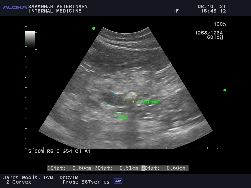
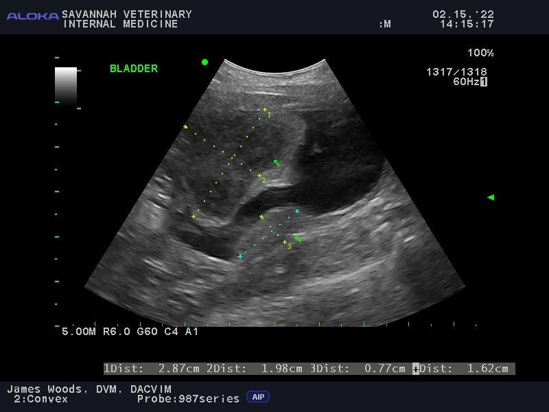
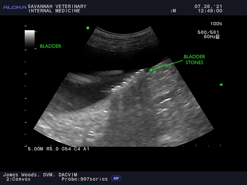
Urinary bladder
Urinary bladder disorders are commonly seen in dogs and cats. A variety of diseases can be detected with ultrasound, including cancer, inflammation, scarring (fibrosis), stones and blood clots. Ultrasound is also very useful for confirming correct placement of urinary catheters that may placed to assist a patient who is having trouble urinating.
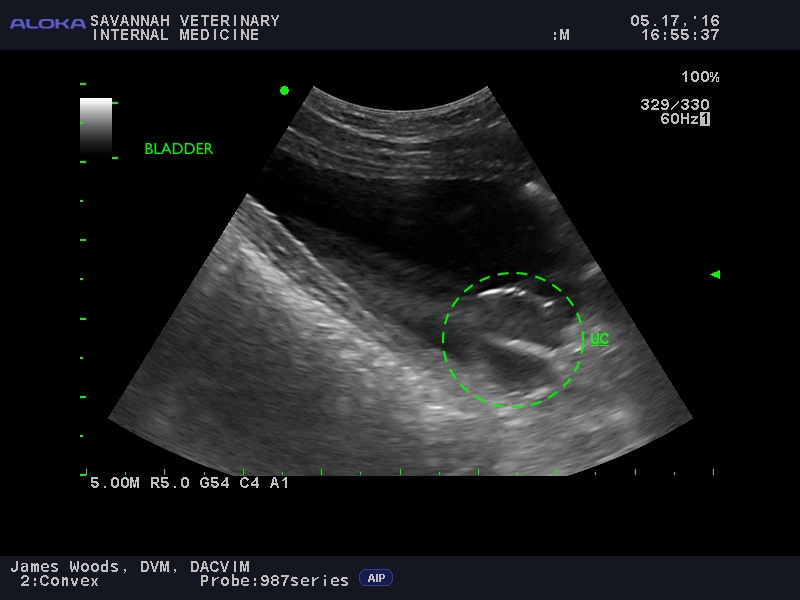
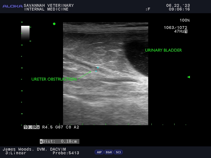
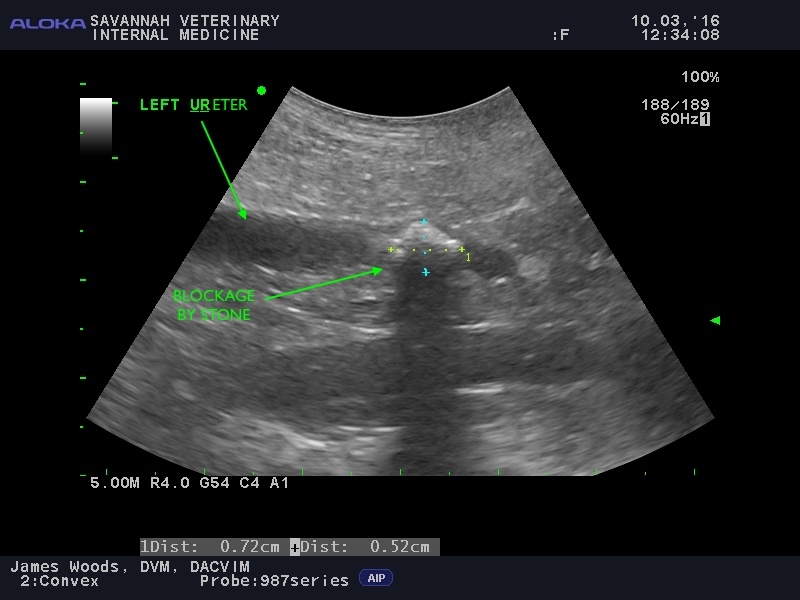
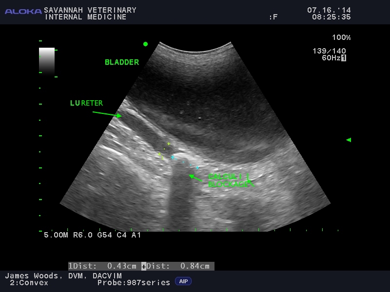
Ureters
The ureters are small tubes that connect the kidneys to the urinary bladder. Urine produced in the kidneys passes down these tubes to fill the urinary bladder. The ureters are so small that normally they cannot be seen with ultrasound. The ureters become visible when they dilate due to obstruction from tumors, inflammation, infection or small kidney stones that did not successfully pass all the way into the urinary bladder.
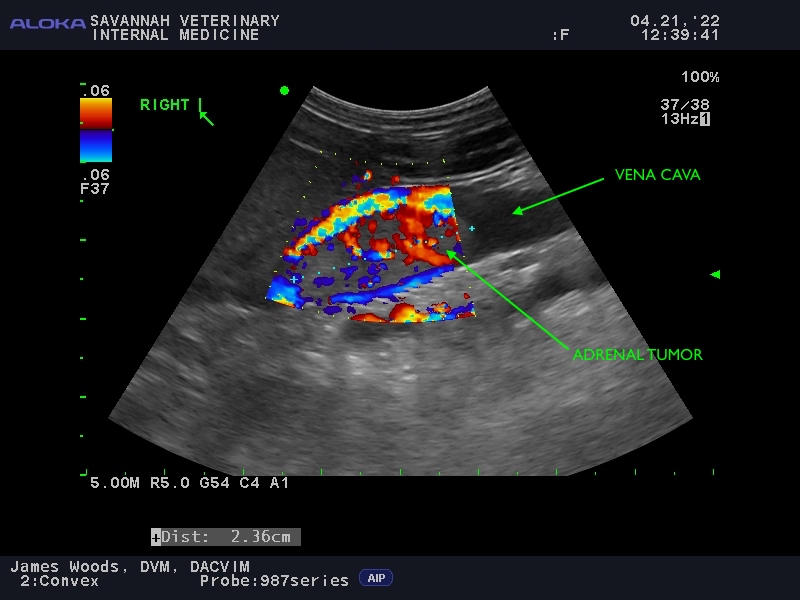
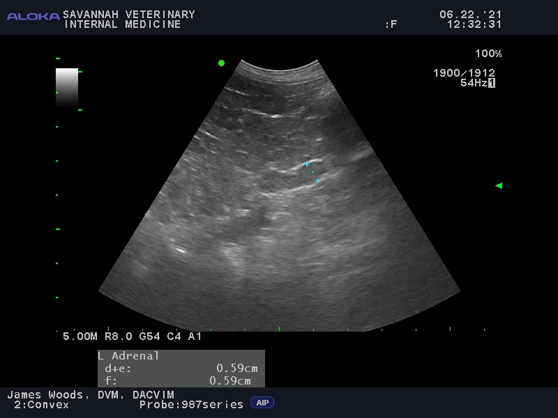
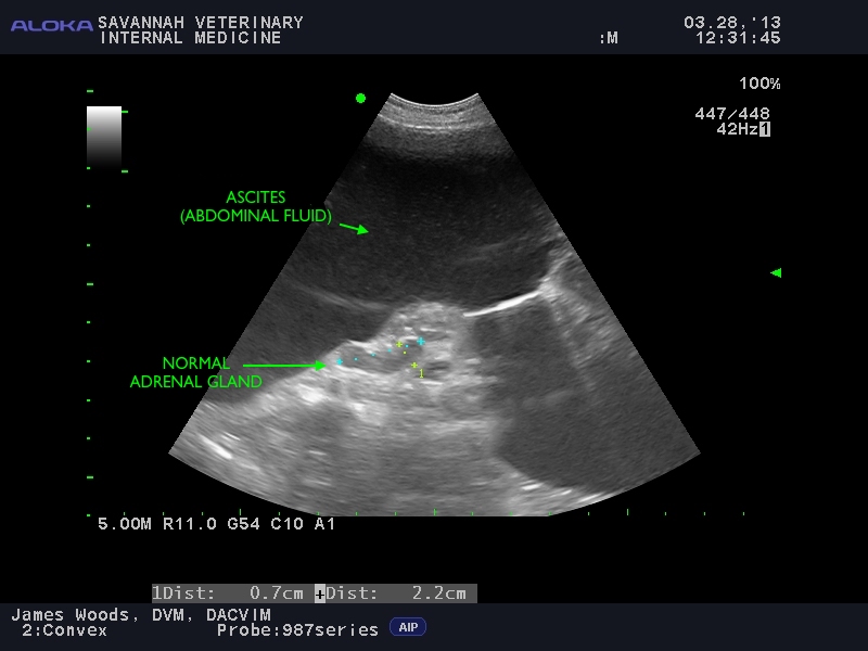
Adrenal glands
The adrenal glands are two small peanut-shaped glands that are located next to each kidney. These glands produce important hormones that control metabolism, immune functions, blood pressure and stress (adrenaline, “fight or flight” response). Normal adrenal glands are too small to be seen with X-rays, but ultrasound can easily visualize normal and diseased adrenal glands, in both dogs and cats. Diseases that affect the adrenal glands include cancer, Cushing’s disease, pheochromocytoma, Addison’s disease and hyperaldosteronism.

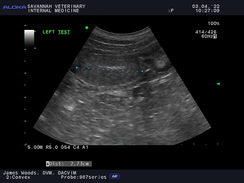
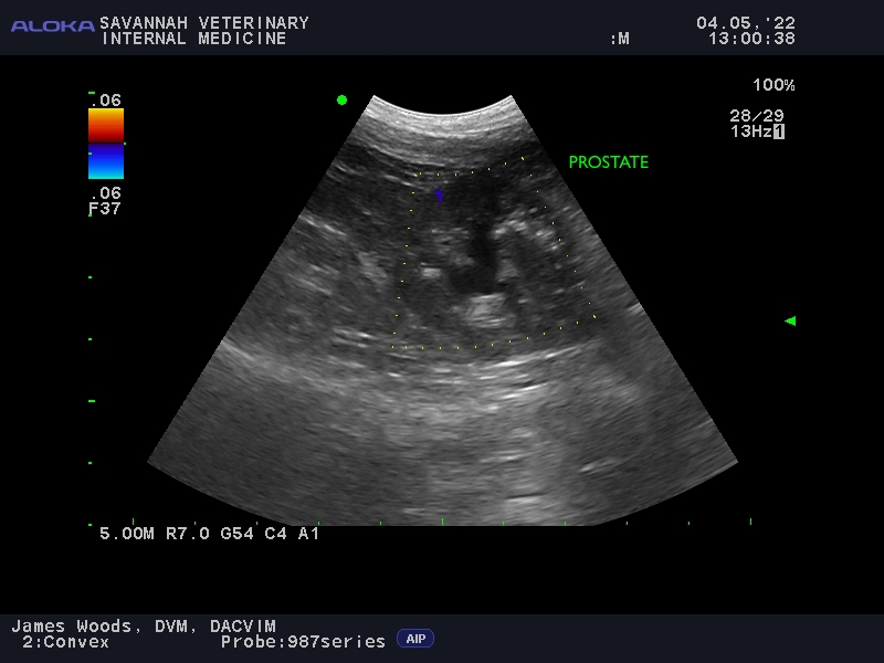
Reproductive
Spaying and neutering (castration) not only prevents pregnancy, but it also reduces the risk of reproductive diseases such as sexually-transmitted diseases (Brucellosis), mammary (breast) cancer, testicular cancer, pyometra (infection within the uterus), and prostatitis (infection of the prostate). Unfortunately, even male dogs that are neutered are at risk of developing prostate cancer.
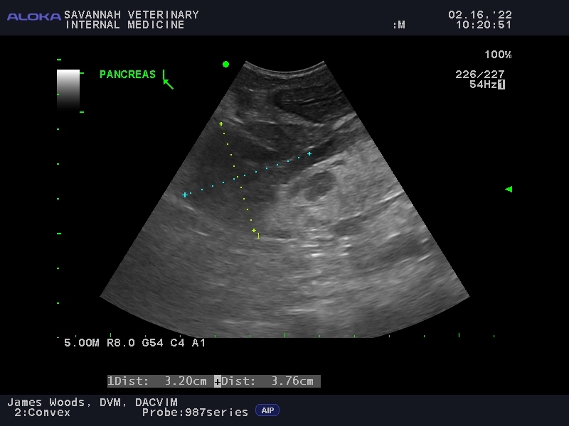
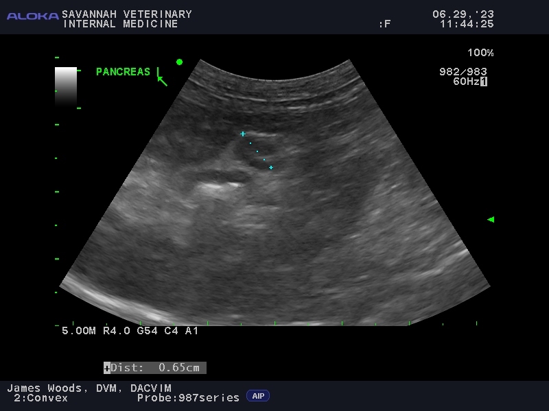
Pancreas
The pancreas produces insulin (when this is low, diabetes mellitus occurs) and digestive enzymes which mixes with food in the intestines (when this is low, a disease called exocrine pancreatic insufficiency — aka EPI — occurs). Although these diseases do not usually show up on ultrasound, a variety of diseases do, including pancreatitis (benign inflammation of the pancreas) and pancreatic cancer (e.g. insulinoma).
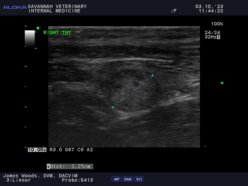
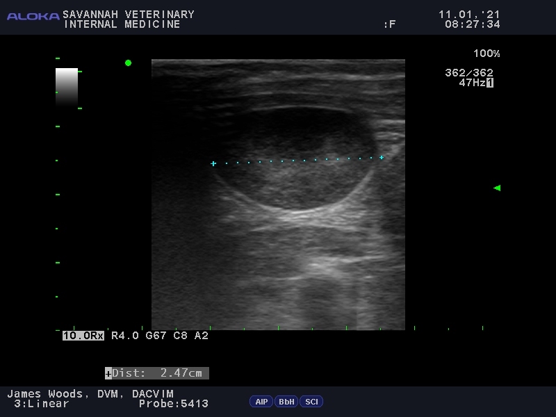
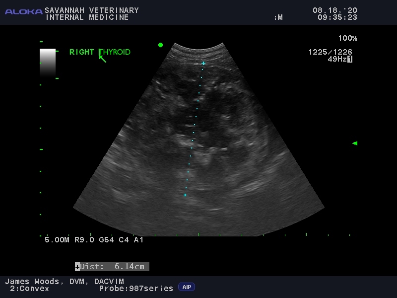
Thyroid
The thyroid glands are located under the skin in the neck and are responsible for regulating metabolism. Ultrasound is very useful in detecting thyroid diseases, including benign adenomas and malignant cancer.
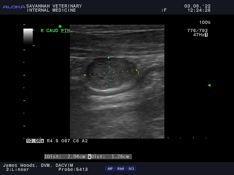
Parathyroid
The parathyroid glands — located just under the skin in the neck next to the thyroid gland— regulate calcium in the body. Disorders of the parathyroid glands (primary or secondary hyperparathyroidism) cause an imbalance in the body’s calcium concentration. Ultrasound excels at detecting swelling and masses in these small glands.


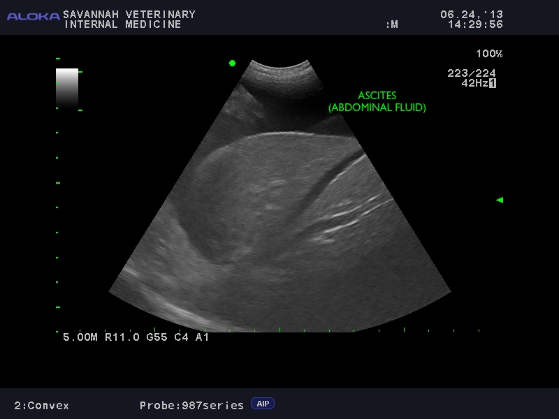
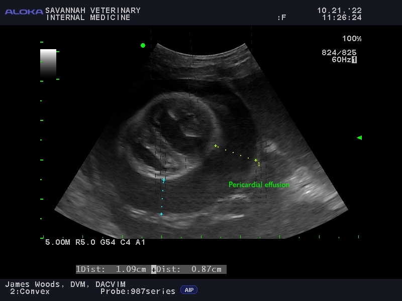
Abnormal fluid
Abnormal fluid can accumulate in many places in the body. Three of the most common locations we identify with ultrasound include in the abdominal cavity (ascites), in the chest cavity (pleural effusion) and around the heart (pericardiac effusion). Usually serious disorders cause fluid accumulation and sampling the fluid with a needle for analysis is an important test to identify the cause.

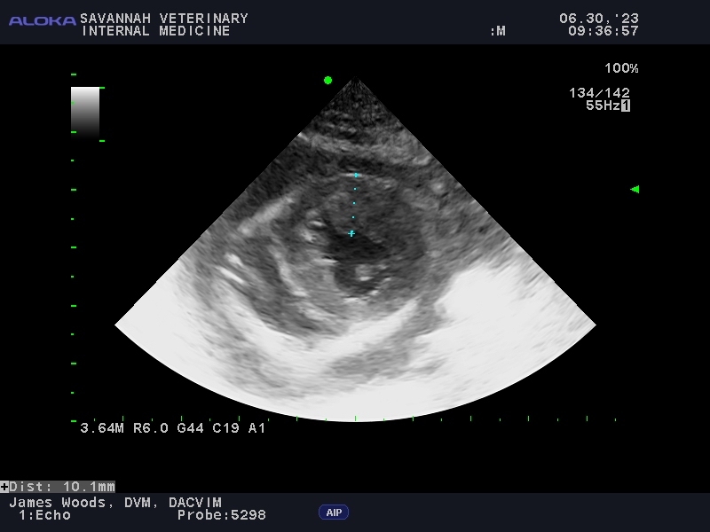
Heart
Heart diseases are easily diagnosed with ultrasound. Common diseases encountered include cardiomyopathy (dilated, hypertrophic), myxomatous mitral valve disease, congenital heart diseases (pulmonic stenosis, aortic stenosis), heartworm disease, pulmonary hypertension and heart cancer. Some of these can result in abnormal fluid accumulation within the sac that surrounds the heart (pericardial effusion).

Lung
In many situations, X-rays are valuable in detecting abnormal masses and cancers within the lungs. However, lung masses and cancers that are located on the “edge” of the lungs are easily viewed with ultrasound. Unlike X-rays, ultrasound can then be used to direct a needle through the chest wall and sample (aspirate) these lung masses for diagnosis.


We are experts at veterinary ultrasound
If it has been recommended that you pet needs an ultrasound, contact us and we can help. Now you know what we are looking for, and what may, or may not be found.