When our beloved dogs and cats are under the weather, figuring out what’s wrong can feel like a mystery. Fortunately, veterinary medicine has powerful imaging tools to look inside our furry companions, helping veterinarians diagnose disease with greater speed and accuracy. While you may be familiar with these tests in human medicine, there are some unique aspects when it comes to our pets.
This blog explores the common imaging tools your veterinarian might recommend: radiographs (X-rays), ultrasound, CT scans, and MRI, and what they can tell us about your pet’s health.
Radiographs (X-rays): The Skeletal Snapshot
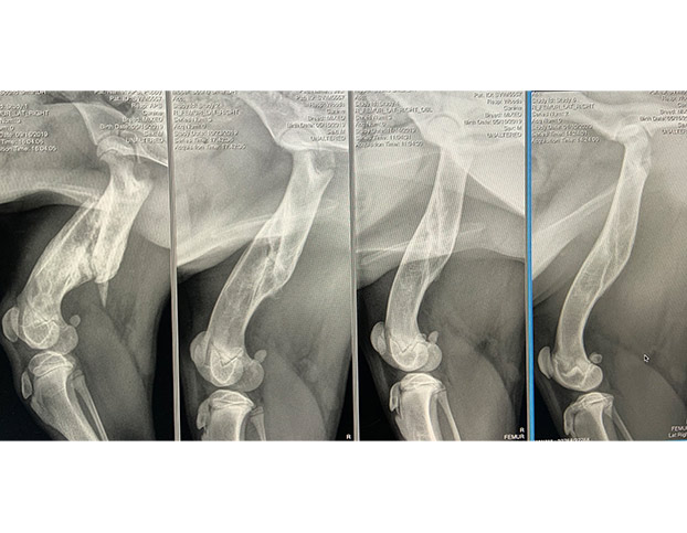
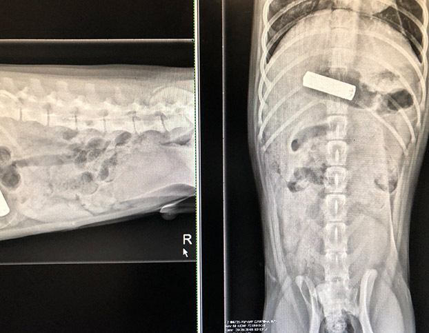
What they are: Radiographs, commonly known as X-rays, are one of the most widely used diagnostic tools in veterinary medicine. They provide a two-dimensional image of your pet’s internal structures.
What it excels at:
- Excellent for bones: X-rays are the best tool for identifying fractures, arthritis, and other bone abnormalities.
- Visualize foreign objects: Swallowed toys, rocks, metal objects or other foreign bodies in the digestive tract are often clearly visible on X-rays
- See organ size and shape: While not providing detailed internal structure of organs, X-rays can reveal changes in the size, shape, and position of organs like the heart, lungs, liver, and kidneys. They can help detect conditions like an enlarged heart or fluid in the lungs.
What to be aware of:
- Limited soft tissue detail: X-rays don’t provide detailed images of the internal structure of soft tissues and organs like they do bones.
- Superimposition: Because it’s a 2D image, structures can overlap, potentially hiding underlying issues.
- Cannot visualize fluid-filled or gas-filled structures well internally: While they can show the presence of fluid or gas in body cavities, they aren’t ideal for examining the details within fluid-filled cysts or the structure of gas-filled intestines.
Ultrasound: Seeing in Waves
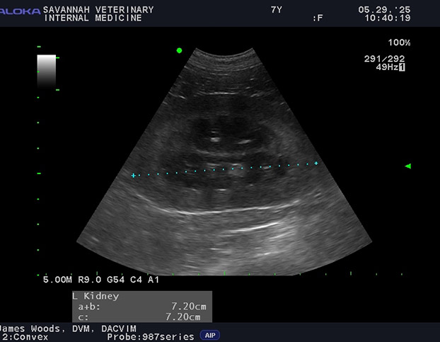
What it is: Ultrasound uses sound waves to create real-time images of your pet’s internal organs and structures.
What it excels at:
- Excellent soft tissue detail: Ultrasound provides detailed images of the internal architecture of organs like the liver, kidneys, spleen, bladder, and reproductive organs.
- Visualize fluid and abnormal tissues: It’s ideal for detecting free fluid in body cavities, cysts, abscesses, and changes in organ texture that might indicate disease like tumors.
- Real-time imaging: Allows veterinarians to observe organ movement, blood flow (with Doppler ultrasound), and guide procedures like needle biopsies.
- Non-invasive and no radiation: Ultrasound is very safe for patients and veterinary staff, and is safe for pregnant animals and for frequent monitoring of disease progression.
What to be aware of:
- Sound waves don’t travel through air or bone: This makes ultrasound less useful for examining the lungs (unless there’s fluid accumulation or a lung tumor) or structures hidden by bone, like the brain, spinal cord and pelvic canal.
- Operator dependent: The quality and interpretation of ultrasound images rely heavily on the skill and experience of the person performing the scan. Everyone can look at an X-ray or a CT scan image and see the same thing; this is not the case with ultrasound: experience matters.
- Patient cooperation is helpful: While we do not need sedation or anesthesia for the ultrasounds we perform, a pet that is very wiggly can make obtaining good images challenging.
CT Scan (Computed Tomography): Detailed Cross-Sections
What it is: A CT scan (“CAT scan”) uses a series of X-ray images taken from multiple angles to create detailed cross-sectional slices of your pet’s body. These slices can then be reconstructed by a computer to form 3D images.
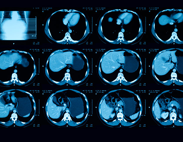
What it excels at:
- Excellent for bony structures and complex anatomy: CT provides much more detail of bone than traditional X-rays and is great for examining complex areas like the skull, spine and nasal cavity.
- Visualize internal organs in detail: While ultrasound is great for soft tissue, CT can provide more comprehensive views of organs, especially when looking for masses or structural changes.
- Identify subtle lesions: The cross-sectional images can reveal small abnormalities that might be missed on traditional X-rays and is very helpful for looking for metastatic cancer in the lungs.
What to be aware of:
- Involves higher doses of radiation than traditional X-rays: If a scan is considered important to do for the health of your pet, the benefits usually outweigh the risks, but it’s a consideration.
- Requires general anesthesia: Pets must remain perfectly still for the scan, necessitating general anesthesia or deep sedation.
MRI (Magnetic Resonance Imaging): The Soft Tissue Specialist
What it is: MRI is an advanced imaging technique that uses a strong magnetic field to create highly detailed images of your pet’s soft tissues, particularly the brain, spinal cord, muscles, and ligaments.
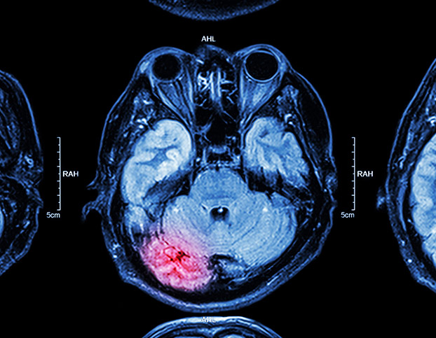
- Superior soft tissue detail: MRI is the best tool for visualizing and evaluating the brain, spinal cord, nerves, muscles, tendons, and ligaments.
- Detects subtle changes in tissue composition: Can identify inflammation, tumors, fluid accumulation, and other abnormalities within soft tissues with high accuracy.
- Ideal for neurological conditions: The preferred imaging technique for diagnosing conditions affecting the brain and spinal cord, such as herniated discs, tumors, and inflammation.
- Does not image bone well: Bone appears dark on MRI, making it less useful for evaluating bone structure itself compared to X-rays or CT.
- Requires general anesthesia: Pets must be under general anesthesia to remain completely still for the duration of the scan, which can be longer than a CT scan.
- More expensive and less available: MRI equipment is costly and not widely available.
- Cannot be used in pets with certain metal implants: The strong magnetic field can interfere with or heat up some metallic implants.
Special Considerations for Pets vs. People
One of the most significant differences when your pet undergoes advanced imaging like CT or MRI compared to a person is the necessity of sedation or general anesthesia. While humans can understand the need to lie still and hold their breath, pets cannot. Any movement during these scans can blur the images, making them non-diagnostic. Therefore, to obtain high-quality images crucial for accurate diagnosis, pets are typically heavily sedated or placed under general anesthesia. This requires careful pre-anesthetic evaluation, including blood work and a physical examination, to ensure your pet is healthy enough for the procedure. Another consideration is the cost. Advanced imaging tools like CT and MRI require specialized equipment, making them more expensive than radiographs or ultrasound. Finally, unlike humans who can communicate their symptoms and discomfort, veterinarians rely heavily on physical examination findings, your observations of your pet’s behavior, and the information gained from imaging to piece together a diagnosis. Diagnostic imaging plays a vital role in this puzzle, allowing vets to visualize what they cannot see or feel externally.Contact Us
In conclusion, veterinary imaging tools are invaluable to look beyond the surface and diagnose a wide range of conditions in our beloved dogs and cats. While the procedures may differ slightly from those in human medicine, the goal is the same: to gather the information needed to provide the best possible care for your furry family member. If you have questions about these tests, or think that your pet may benefit from imaging due to their symptoms, contact us we can help determine the next best step for your pet’s health journey.
Author:
James Woods DVM, MS, DACVIM (SAIM)
Ph: (912) 721-6410
Contact Us